Macromastia is defined as a medical condition in which the breasts contain abnormally excessive glandular, fatty, or fibrous tissue, causing significant physical and psychological burden.
Clinically, the term breast hypertrophy is used interchangeably, though macromastia typically refers to cases where breast weight causes measurable functional impairment. The British Association of Aesthetic Plastic Surgeons (BAAPS) recognises breast reduction as one of the most consistently high-satisfaction procedures performed in the UK, reflecting how profoundly this large breast condition affects quality of life.
This article draws on current research, including findings published in 2026, to explain the causes, symptoms, diagnostic approaches, and treatment options available to patients in the UK.
What causes macromastia?
Macromastia is not simply a consequence of weight gain. The condition involves a complex interplay of hormonal, genetic, and systemic factors that drive abnormal breast tissue growth beyond normal variation.
Hormonal pathways
Research published in january 2026 identifies increased expression of aromatase (CYP19A1) and heightened prolactin sensitivity as key hormonal mechanisms in macromastia. Aromatase is the enzyme responsible for converting androgens into oestrogen within breast tissue. When this enzyme is overexpressed locally, oestrogen levels within the breast rise disproportionately, stimulating excessive tissue proliferation. Prolactin dysfunction compounds this effect by further promoting glandular growth. Together, these endocrine pathways explain why macromastia can develop or worsen during puberty, pregnancy, and hormonal therapy, even in women without significant overall weight gain.
Genetic and systemic contributors
Genetic mutations, including mutations in the PTEN tumour suppressor gene, have been implicated in some cases of macromastia. PTEN mutations disrupt normal cell growth regulation, which can manifest in breast tissue as uncontrolled proliferation. Autoimmune conditions, particularly systemic lupus erythematosus, are also associated with the condition. The mechanism here likely involves inflammatory cytokines that alter local tissue sensitivity to hormonal signals. These associations mean that a thorough medical history is an essential part of any clinical assessment.
Obesity and body mass index
Approximately two-thirds of women with macromastia are obese, indicating a strong correlation between body mass index and excessive breast tissue volume. This correlation matters clinically because adipose tissue itself contains aromatase, meaning that higher overall body fat can amplify local oestrogen production within the breast. However, obesity alone does not cause macromastia. Many patients with a healthy body mass index develop the condition, confirming that hormonal and genetic factors operate independently of weight.
Macromastia vs gigantomastia
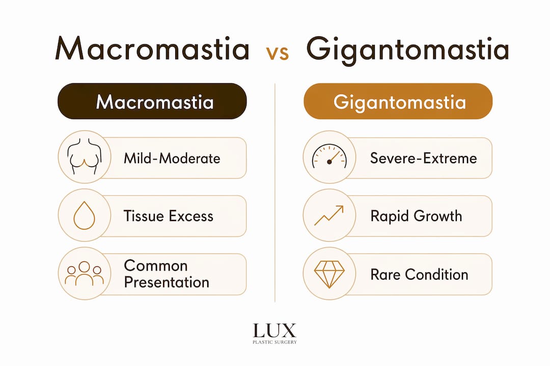
Macromastia and gigantomastia both describe abnormally large breasts, but gigantomastia refers to the most extreme end of the spectrum, where breast tissue growth is rapid, severe, and often associated with pregnancy or medication triggers. Gigantomastia is rarer and may require more urgent surgical intervention. Macromastia, by contrast, describes a broader range of cases where excess tissue causes functional impairment without necessarily reaching the extreme volumes seen in gigantomastia.
Pro Tip: If you notice rapid breast enlargement over a period of weeks rather than months, seek a clinical assessment promptly. Rapid growth can indicate gigantomastia or an underlying systemic trigger that requires investigation before any surgical planning begins.
What symptoms and complications does macromastia cause?
Macromastia causes a range of physical and psychological symptoms that worsen progressively if left unmanaged. The severity of symptoms does not always correlate directly with breast volume. Some patients with moderately enlarged breasts experience debilitating pain, while others with larger volumes report primarily psychosocial distress.
Musculoskeletal pain and postural strain
Chronic pain in the back, neck, and shoulders is the most commonly reported symptom of macromastia. The weight of the breasts shifts the body’s centre of gravity forward, forcing the spine into a compensatory posture. Over time, this altered biomechanics causes muscle fatigue, joint strain, and in some cases, disc compression in the cervical and thoracic spine. Patients frequently report that pain worsens with prolonged standing, physical activity, or wearing a bra for extended periods.

Nerve injury and sensory loss
Persistent breast weight exerts chronic traction on the 4th, 5th, and 6th intercostal nerves, which supply sensation to the nipple and surrounding areolar tissue. This traction can cause permanent stretch injury to these nerves, resulting in partial or complete loss of nipple sensation. The critical clinical point is that this sensory loss may not fully recover even after surgical reduction. Patients should be counselled about this risk before surgery, as it affects both expectation setting and the timing of intervention.
Skin complications
Skin chafing, rashes, and ulceration beneath the breast fold are common in macromastia. The inframammary fold creates a warm, moist environment where skin surfaces rub together, leading to intertrigo, fungal infections, and in severe cases, skin breakdown. Overstretched skin loses its elasticity and becomes prone to excoriation. These dermatological complications are often underreported by patients who regard them as a hygiene issue rather than a medical symptom.
Psychosocial impact
The psychological burden of macromastia is substantial and clinically significant. Patients report difficulties with clothing, avoidance of physical activity, social withdrawal, and reduced self-confidence. The condition affects participation in exercise, which in turn can worsen obesity and the hormonal factors that drive further breast tissue growth. Addressing psychosocial distress is therefore not a secondary concern. It is a core component of the clinical assessment and treatment planning process.
How is macromastia diagnosed and assessed?
Diagnosis of macromastia has historically relied on subjective measures such as bra cup size and patient-reported symptoms. Current clinical practice is moving away from this approach in favour of objective, quantitative assessment tools that improve both diagnostic accuracy and surgical planning.
Traditional vs modern diagnostic methods
| Method | Approach | Limitation or benefit |
|---|---|---|
| Bra cup size | Subjective patient report | Inconsistent across manufacturers; poor clinical reliability |
| Inframammary fold measurement | Physical measurement of fold depth and ptosis | Reproducible; useful for severity classification |
| 3D surface imaging | Digital volumetric modelling of breast tissue | Quantitatively superior; improves surgical outcome prediction |
| Symptom scoring | Standardised questionnaires for pain and function | Captures functional impact; complements objective measures |
A paradigm shift toward volumetric assessment and inframammary fold measurements is now documented in the clinical literature. This shift matters because cup size varies between manufacturers and provides no information about tissue composition or distribution. Volumetric data, by contrast, allows the surgeon to calculate the precise weight of tissue to be removed and to model the expected postoperative result.
3D imaging in clinical practice
3D imaging and digital models provide reliable volumetric measurements that are quantitatively superior to traditional subjective assessments. In practice, this means a patient can view a simulated postoperative result before committing to surgery, which supports informed consent and realistic expectation setting. Inframammary fold measurements complement 3D imaging by providing a physical reference point for surgical incision planning. Together, these tools represent the current standard of care for macromastia assessment in specialist plastic surgery centres.
Severity classification
Macromastia is classified by the volume of tissue exceeding normal breast weight thresholds. Mild macromastia typically involves excess tissue of 200–600 grams per breast. Moderate cases involve 600–1,500 grams, and severe cases exceed 1,500 grams per breast. Gigantomastia is generally defined as excess tissue above 2,000 grams per breast. These thresholds guide surgical planning and help determine whether a patient is likely to meet NHS criteria for funded breast reduction surgery.
Pro Tip: When attending a consultation, bring a record of any physical therapy, pain management, or dermatological treatments you have already tried. Documented conservative treatment history strengthens the clinical case for surgical intervention and supports any NHS funding application.
What treatment options are available for macromastia?
Treatment for macromastia ranges from conservative symptom management to definitive surgical correction. The appropriate approach depends on symptom severity, the patient’s overall health, and their personal goals.
Non-surgical management
Non-surgical treatments address symptoms rather than the underlying cause. They are appropriate for patients with mild macromastia, those who are not yet surgical candidates, or those managing symptoms while awaiting surgery.
- Joint corticosteroid injections and physical therapy are the primary non-surgical interventions for pain relief in macromastia. Corticosteroid injections reduce inflammation in affected joints, while physical therapy addresses postural imbalances and strengthens supporting musculature.
- Specialist bra fitting with structured support reduces the mechanical load on the spine and can provide meaningful short-term pain relief.
- Dermatological treatment of inframammary skin conditions, including antifungal creams and barrier preparations, manages skin complications but does not address their root cause.
- Weight management, where relevant, can reduce the adipose component of breast tissue and lower local oestrogen production via reduced aromatase activity.
- Psychological support, including cognitive behavioural therapy, addresses the psychosocial dimension of the condition and is recommended as part of a multidisciplinary approach.
Non-surgical management does not correct the structural problem. For patients with moderate to severe macromastia, it provides temporary relief while surgical options are considered.
Surgical treatment: reduction mammaplasty
Reduction mammaplasty is the definitive surgical treatment for macromastia. The procedure removes excess glandular tissue, fat, and skin, and repositions the nipple-areola complex to a natural height. It is performed under general anaesthetic and typically takes two to four hours. The result is a smaller, lighter breast that resolves the biomechanical causes of pain and skin complications.
The main surgical steps in reduction mammaplasty are as follows:
- Preoperative marking. The surgeon marks incision lines and the new nipple position with the patient standing, using volumetric data from 3D imaging to guide planning.
- Tissue removal. Excess glandular tissue, fat, and skin are excised. The volume removed is weighed intraoperatively to confirm it matches the preoperative plan.
- Nipple-areola repositioning. The nipple-areola complex is moved to the new position, either on a pedicle of tissue (preserving blood supply and sensation) or, in very large reductions, as a free nipple graft.
- Wound closure. The remaining skin is reshaped and closed, typically leaving an anchor-shaped or vertical scar pattern depending on the technique used.
- Postoperative recovery. Patients wear a supportive surgical bra for six weeks. Most return to light activity within two to three weeks and to full activity within six to eight weeks.
Risks of reduction mammaplasty include scarring, changes in nipple sensation, asymmetry, wound healing complications, and, rarely, partial loss of the nipple-areola complex. Scar management after surgery is an active area of clinical interest. Light therapy for scar reduction is one emerging approach that some patients explore postoperatively to improve scar appearance over time.
For patients considering the full range of breast surgery options, understanding how reduction mammaplasty compares to other procedures is an important part of the decision-making process.
Key takeaways
Macromastia is a medically recognised condition requiring objective clinical assessment and, in moderate to severe cases, surgical correction through reduction mammaplasty to achieve lasting symptom relief.
| Point | Details |
|---|---|
| Hormonal mechanisms are central | Overexpression of aromatase (CYP19A1) and prolactin sensitivity drive abnormal breast tissue growth. |
| Symptoms extend beyond pain | Nerve damage, skin ulceration, and psychosocial distress are all documented complications of untreated macromastia. |
| Objective diagnosis improves outcomes | 3D volumetric imaging and inframammary fold measurement replace cup size as the clinical standard for surgical planning. |
| Non-surgical care manages symptoms only | Physical therapy and corticosteroid injections relieve pain temporarily but do not correct the underlying structural problem. |
| Reduction mammaplasty is definitive | Surgical removal of excess tissue under general anaesthetic provides long-term relief and is associated with high patient satisfaction. |
Professor Hindocha’s perspective on managing macromastia
What strikes me most, having assessed many patients with macromastia over the years, is how long most of them have waited before seeking a formal clinical opinion. By the time they arrive at a consultation, the majority have been managing chronic pain, skin problems, and significant emotional distress for years. They have often been told that their symptoms are simply a consequence of their body shape, rather than a recognised medical condition with established treatment pathways.
The shift toward objective diagnostic tools has changed the quality of these consultations considerably. When a patient can see a volumetric model of their breast tissue and understand precisely how much excess weight they are carrying, the conversation becomes grounded in clinical fact rather than subjective perception. That clarity matters. It helps patients make informed decisions and sets realistic expectations about what surgery can and cannot achieve.
One point I emphasise consistently is the question of nerve injury. Chronic traction on the intercostal nerves is a real and underappreciated risk of delayed treatment. Patients sometimes assume that waiting longer before surgery is the cautious choice. In reality, prolonged nerve traction can result in sensory loss that surgery cannot reverse. Timing matters, and earlier assessment is generally in the patient’s interest.
The other aspect I would highlight is the importance of a multidisciplinary approach. Reduction mammaplasty addresses the structural problem effectively, but patients benefit from physiotherapy, psychological support, and dermatological care as part of a complete treatment plan. Surgery is not the entirety of the solution. It is the central intervention within a broader programme of care.
— Lux
Breast reduction surgery at Lux Plastic Surgery
Lux Plastic Surgery offers breast reduction surgery for patients with macromastia, performed by Professor Sandip Hindocha, GMC-registered Consultant Plastic Surgeon and NHS Clinical Director. Consultations are available at clinics in Bedford, London, and Manchester.

Professor Hindocha uses 3D volumetric assessment and individualised surgical planning to guide each procedure. Patients receive a full preoperative assessment, clear information on risks and recovery, and access to postoperative support. For patients weighing surgical versus non-surgical options, the consultation process covers both pathways in detail. To review the full range of procedures available, visit the Lux Plastic Surgery services page.
This article is for informational purposes only and does not constitute medical advice. Consult a GMC-registered specialist for a clinical assessment.
FAQ
What is macromastia?
Macromastia is a medical condition defined by abnormally excessive breast tissue causing physical symptoms such as back pain, nerve injury, and skin complications. It is also referred to clinically as breast hypertrophy.
What causes macromastia to develop?
The primary causes include overexpression of aromatase (CYP19A1), heightened prolactin sensitivity, genetic mutations such as PTEN, autoimmune conditions including lupus, and a strong association with obesity.
Can macromastia be treated without surgery?
Non-surgical treatments including physical therapy and corticosteroid injections manage pain temporarily but do not correct the underlying excess tissue. Reduction mammaplasty is the only definitive treatment.
Does macromastia qualify for NHS funding?
NHS funding for breast reduction surgery depends on documented functional impairment, evidence of conservative treatment failure, and meeting local clinical commissioning criteria. A specialist assessment is required to determine eligibility.
What is the recovery time after reduction mammaplasty?
Most patients return to light daily activity within two to three weeks and resume full physical activity within six to eight weeks. A supportive surgical bra is worn for approximately six weeks postoperatively.